
Why Is a Lead Apron Used for X Ray Protection?

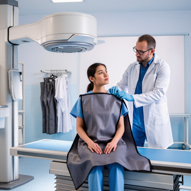
A lead apron is used for X-ray protection because it can reduce exposure to scattered radiation. During an X-ray examination, the machine directs a controlled beam through the body. Some radiation changes direction after interacting with tissues. This scattered radiation may reach nearby organs, staff, or a patient’s unexamined body areas. A properly fitted lead apron can absorb part of that scattered energy.

The material is usually a flexible composite containing lead or another radiation-attenuating substance. It is designed to block or weaken X-rays, not to make exposure disappear. That distinction matters. A lead apron does not replace distance, shielding screens, careful positioning, or shorter exposure times. It also should never be placed in the primary beam unless specifically approved for a procedure. Wrinkles and cracks may reduce its protective performance.

Medical imaging professionals assess protection according to the examination, equipment, and exposure risk. A radiographer may check the apron’s condition, hang it correctly, and keep it away from unnecessary pressure. In fluoroscopy rooms, staff often use aprons, thyroid collars, ceiling shields, and mobile barriers together. Patients may receive shielding when it supports safe practice, though modern guidance does not require routine shielding for every examination. Practices can vary.

The explanation is not perfectly simple. More protection is not always better if it interferes with positioning or causes repeat images. The safest approach combines professional judgment, current clinical guidance, and well-maintained equipment. That balance helps protect people without creating a false sense of security.
Why Is a Lead Apron Used for X Ray Protection?

What X Rays Are and How They Interact With the Human Body

Why Is a Lead Apron Used for X Ray Protection?

X rays are high energy photons, not visible light. They can travel through soft tissue and create diagnostic images on a detector. Their interaction depends on tissue thickness, density, and photon energy. Bone absorbs more X rays than muscle, so it appears brighter. Air absorbs very little, which makes the lungs look darker.

Inside the body, X rays may transfer energy to atoms. This process can produce ionization and damage cellular DNA. Most exposure is low, but repeated or unnecessary exposure deserves careful control. Scatter also matters. When an X ray changes direction after meeting tissue, it can reach nearby staff or body areas.

A lead apron helps by absorbing and weakening scattered radiation. Its dense material reduces the amount reaching sensitive organs. It is not a complete barrier. It cannot remove all risk, and poor positioning can leave gaps. In a radiology room, distance, short exposure time, and correct equipment settings remain essential. Professional guidance usually supports shielding when it is appropriate to the procedure and local policy. Routine use is not always required, especially when it interferes with imaging or protects an area already outside the beam. That point can feel counterintuitive. A thicker apron is not automatically better. Careful planning often protects people more effectively than simply wearing extra material.

Why Repeated X Ray Exposure Requires Protective Measures

Why Is a Lead Apron Used for X Ray Protection?

Why Repeated X Ray Exposure Requires Protective Measures

X rays pass through the body to create an image, but some energy scatters around the patient. Repeated exposure can increase long-term radiation risk, even when each examination uses a low dose. The risk is usually small, but it is not zero. That distinction matters.

A lead or lead-equivalent apron helps reduce scattered radiation reaching the torso. It does not make someone invulnerable, and it should never replace distance, shielding, or careful positioning. During fluoroscopy, staff can step behind a protective barrier, move away from the beam, and avoid standing close to the patient’s tube side. Small changes can reduce exposure.

Fit matters too. An apron should cover the front and sides of the body without restricting movement. It should not be folded, because creases may damage its protective layers. Regular visual and radiographic inspections can reveal hidden cracks. Wear monitoring may also help workers understand their exposure patterns.

In a busy imaging room, routines can become automatic. A misplaced apron or unnecessary time near the table is easy to overlook. That is where professional judgment remains important. Each examination should have a clear clinical purpose, and protective measures should match the actual working conditions. Even experienced teams need reminders. Mistakes happen. Habits should be checked.

Why Is a Lead Apron Used for X-Ray Protection?

Approximate reduction of scattered X-ray radiation provided by common lead-equivalent apron thicknesses at about 100 kVp

Repeated X-ray exposure can accumulate over time and increase the probability of radiation-related effects. Lead aprons reduce exposure to scattered radiation reaching radiosensitive organs, with thicker lead-equivalent material generally providing greater attenuation. The actual protection depends on photon energy, fit, apron condition, and exposure geometry. Aprons should be used together with minimized exposure time, increased distance, and appropriate shielding; they are not designed to stop the primary X-ray beam.

Data shown are approximate reference values for typical protective materials at approximately 100 kVp. Attenuation varies with X-ray spectrum, beam angle, and garment construction.

How Lead Aprons Reduce Scattered Radiation

Why Is a Lead Apron Used for X Ray Protection?

During X-ray imaging, the patient becomes the main source of scattered radiation. When the beam strikes tissue, photons change direction and spread around the room. This scatter is weaker than the primary beam, but repeated exposure still matters. NCRP Report No. 160 identified medical imaging as the largest source of artificial radiation exposure in the United States. A lead apron forms a dense barrier around the torso. It absorbs or weakens many scattered photons before they reach sensitive organs.

How Lead Aprons Reduce Scattered Radiation

Protection depends on thickness, energy, fit, and position. IAEA guidance reports that suitable protective garments can reduce scattered radiation by more than 90% in many diagnostic settings. However, this figure is not universal. A 0.25 mm lead-equivalent apron may perform differently from a 0.50 mm garment at higher tube voltages. The apron works best with distance, shielding, and shorter exposure time. The ICRP recommends an occupational effective dose limit of 20 mSv per year, averaged over five years. An apron does not make unlimited exposure safe. It is one control, not the entire system. That detail is easy to overlook.

Tips: Keep the front panel closed and inspect it for folds or cracks. Store the apron flat or on a supported hanger. Never use damaged protection without inspection. Step back during fluoroscopy whenever practical. Distance can protect better than heavier clothing. Review dosimeter readings regularly; they may reveal habits that feel safe but are not.

Why Is a Lead Apron Used for X Ray Protection? - How Lead Aprons Reduce Scattered Radiation

Protection Dimension What It Means Relevant Data or Fact Practical Implication
Primary purpose Reduce occupational exposure to scattered X-ray radiation. Scatter is produced when the primary X-ray beam interacts with the patient or another object. Aprons are mainly intended for staff or others who must remain near the patient during an examination.
How lead works Lead attenuates X-rays through its high density and high atomic number. The amount of attenuation depends on lead-equivalent thickness, photon energy, beam angle, and coverage. Thicker or higher lead-equivalent material generally provides greater attenuation but also increases weight and stiffness.
Common apron ratings Lead equivalence describes the shielding performance compared with a specified thickness of lead. Common nominal ratings include 0.25 mm, 0.35 mm, and 0.50 mm lead equivalent. The appropriate rating should be selected according to the procedure, workload, X-ray energy, and local radiation-safety guidance.
Scattered radiation Scatter is less predictable than the primary beam because it depends on patient size, beam direction, field size, and examination geometry. In many diagnostic procedures, the patient is the principal source of scatter to personnel. Standing away from the patient and out of the primary beam remains essential, even when an apron is worn.
Protection of vital organs An apron can shield covered parts of the torso from incident scattered radiation. Protection is limited to the areas covered by the apron; the head, neck, arms, and legs may remain exposed. Correct sizing, full front coverage, and overlap at the sides are important for effective use.
Pregnancy considerations A lead apron may provide additional shielding when a pregnant worker must be near an X-ray source. Declared pregnant workers should follow facility policy, use dosimetry when required, and minimize exposure by time, distance, and shielding. An apron is a supplement to radiation-control measures, not a replacement for safe work practices.
Distance and positioning Radiation intensity from a point source decreases approximately according to the inverse-square relationship. Doubling the distance from the source can reduce exposure rate to approximately one-quarter, when other conditions remain comparable. Use the greatest practical distance and stand at an appropriate angle to the beam and patient.
Primary-beam limitation Protective aprons are not designed to justify intentional exposure to the direct primary beam. The primary beam has much higher intensity than typical scattered radiation and should always be avoided. Never place any body part in the primary beam, even when wearing protective clothing.
Inspection and storage Repeated folding can create cracks or defects in lead-based protective materials. Aprons should be stored flat or on a suitable hanger and inspected according to facility procedures. Damaged or visibly cracked protective garments should be removed from service until evaluated.
Best-practice summary Effective radiation protection combines engineering controls, administrative controls, and personal protective equipment. The core principles are time, distance, shielding, beam restriction, and appropriate monitoring. Use a lead apron only when a person must remain near the source, and always follow applicable local regulations and radiation-safety protocols.

Note: Lead-equivalent ratings and inspection requirements should be verified against the applicable national standard, facility policy, and manufacturer documentation.

When Lead Aprons Are Used During X Ray Procedures

Why Is a Lead Apron Used for X Ray Protection?

When Lead Aprons Are Used During X Ray Procedures

A lead apron reduces exposure from scattered X rays. Scatter forms when the beam meets tissue, equipment, or the imaging table. It is mainly used by people who must remain near the patient. Radiographers, physicians, nurses, and assistants may wear one during fluoroscopy or portable examinations. Patients may receive one only when it will not interfere with imaging.

The apron is most useful when staff cannot leave the room or stand behind a barrier. This often happens during image-guided procedures, where exposure may continue for several minutes. It should cover the torso without restricting movement. Thyroid shields, protective eyewear, distance, and ceiling-mounted screens can provide added protection. The primary beam should not strike the apron unless the procedure requires it. Lead protection cannot replace careful positioning or controlled exposure settings.

In clinical practice, aprons need regular checks for cracks, creases, and poor fit. Damage can create unprotected areas. They should hang flat, never folded over a chair. That detail is easy to overlook. Aprons are unnecessary for every examination. During routine radiography, staff usually leave the room while the patient remains still. Local radiation-safety guidance should decide who wears protection and when. I would not treat an apron as complete protection; time, distance, shielding, and sound technique must work together.

Limitations and Proper Handling of Lead Aprons

Why Is a Lead Apron Used for X Ray Protection?

Lead aprons reduce scatter exposure when staff must remain near a patient during fluoroscopy. Their performance depends on lead equivalence, beam energy, fit, and positioning. They do not block every radiation pathway. ICRP Publication 103 recommends an occupational effective dose limit of 20 mSv per year, averaged over five years. No single year should exceed 50 mSv. NCRP Report 168 stresses optimization through time, distance, shielding, and personal monitoring.

A lead apron is not a substitute for positioning. Standing on the image-receptor side usually reduces scatter compared with standing near the X-ray tube. Keep hands outside the primary beam. Use ceiling-suspended shields and table-mounted curtains when available. Common aprons provide 0.25 or 0.5 mm lead equivalence, but attenuation changes with tube voltage and exposure angle. Coverage can be incomplete. One uncomfortable mistake is treating a heavy apron as permission to stay close.

Handling affects protection. Inspect the apron for tears, hardened areas, or visible creases under local quality-control procedures. Never fold it. Store it vertically or on a supported rack. Periodic fluoroscopic inspection can reveal internal damage that visual checks miss. The IAEA recommends documented inspection programs for protective garments, while NCRP guidance supports replacing damaged equipment promptly. Staff should also avoid hanging aprons over sharp edges or chairs. Small habits matter. Personal dosimeters still matter. A lead apron may reduce dose, but it cannot correct poor workflow, unnecessary exposure time, or careless positioning.